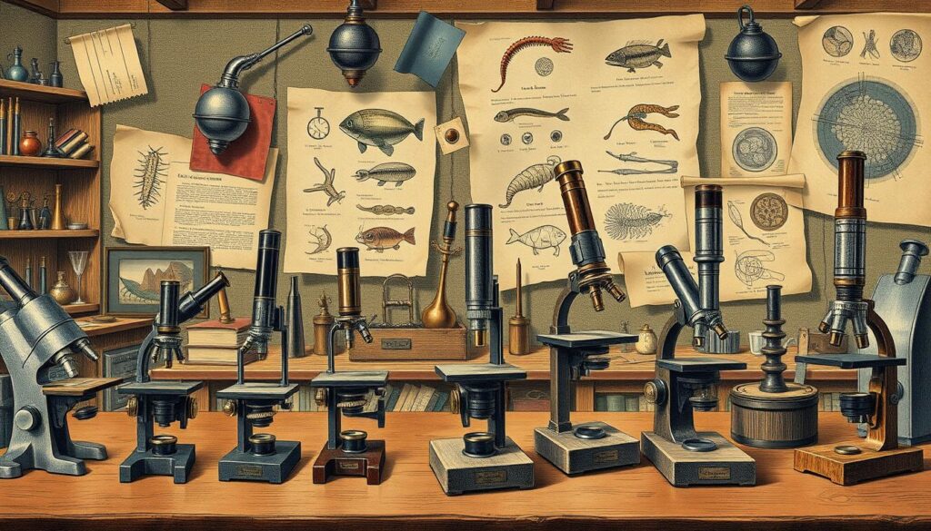
The compound microscope was invented around 1590. It’s a key tool in biology, medicine, and research. Knowing about microscope parts and functions is crucial for using them well.
Since the 16th century, microscopes have greatly helped science. They have parts like objective lenses and eyepiece lenses. These work together to magnify and show clear images of specimens.
Microscope functions like magnification and illumination are key for detailed looks. The simplest microscope, a magnifying glass, offers about 10x magnification. Compound microscopes have eyepiece lenses of 10x or 15x power.
They also have 3 to 4 objective lenses. These lenses have common magnifications of 4x, 10x, 40x, and 100x. This setup allows for high magnification and clear images.
Parts like condenser lenses and Abbe condenser lenses are key for clear images at high magnification. The microscope’s functions, like the diaphragm and rack stop, help control light and movement. Knowing about these parts and functionsis important for using and maintaining the microscope.
Key Takeaways
- Microscopes have been in use since the 16th century, significantly impacting scientific research and discovery.
- Understanding microscope parts, such as objective lenses and eyepiece lenses, is essential for effective use.
- Microscope functions, including magnification and illumination, are critical for detailed examination of structures.
- The combination of microscope parts enables high magnification and clear images.
- Regular maintenance of microscope parts is vital for optimal performance.
- Microscope parts, such as condenser lenses and Abbe condenser lenses, are essential for achieving good resolution at high magnification.
Introduction to Microscopes and Their Importance
Microscopes help us see tiny things like cells and microorganisms. The first microscope was made in 1590 by Hans and Zacharias Jansen. Since then, they’ve become key in biology, medicine, and education.
Today, we have many types of microscopes. Each has special features and uses. Let’s look at some common ones:
- Light microscopes use visible light to show specimens.
- Electron microscopes create images with electrons.
- Fluorescence microscopes use dyes to highlight certain parts.
We’ll dive into how magnification works and the basics of microscopes. Knowing about different microscopes and their uses is vital. This knowledge will be covered in more detail next.
| Type of Microscope | Magnification | Application |
|---|---|---|
| Light Microscope | up to 1000x | Biology, Medicine, Education |
| Electron Microscope | up to 100,000x | Industrial and Medical Research |
| Fluorescence Microscope | up to 1000x | Biology, Medicine, Research |
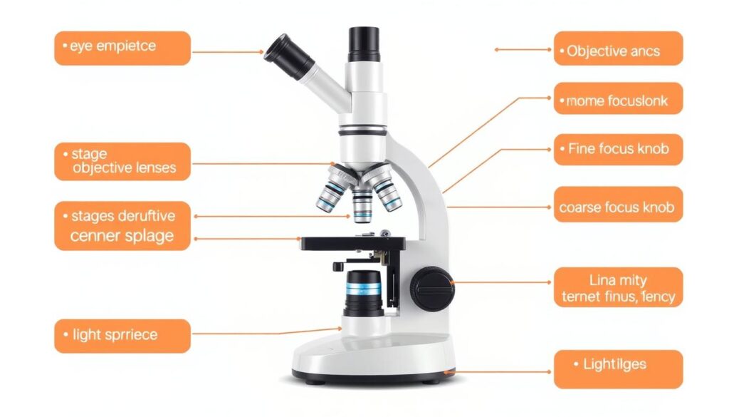
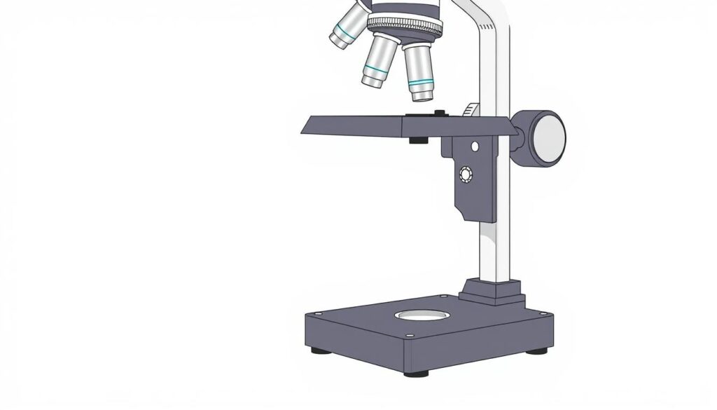
Understanding the Basic Structure of a Microscope
A microscope has several key parts, like the eyepiece, objective lens, stage, condenser, and illuminator. These parts work together to show a magnified view of a specimen. The design of the microscope helps us see tiny details clearly, which is vital in science.
The eyepiece and objective lens are key for magnifying the image. The eyepiece has lenses that can magnify up to 30x, while the objective lens can go up to 100x. The stage and condenser are also important, helping to place the specimen and focus the light.
- Eyepiece: magnifies the image
- Objective lens: gathers light from the specimen
- Stage: holds the specimen in place
- Condenser: focuses the light on the specimen
- Illuminator: provides light for the microscope
Knowing how a microscope works is crucial for making accurate observations. By understanding each part’s role, users can get the best results from their microscope.
| Microscope Component | Function |
|---|---|
| Eyepiece | Magnifies the image |
| Objective Lens | Gathers light from the specimen |
| Stage | Holds the specimen in place |
| Condenser | Focuses the light on the specimen |
| Illuminator | Provides light for the microscope |
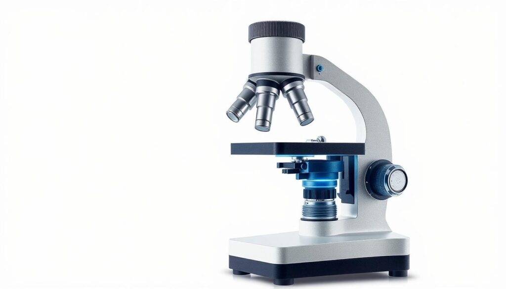
The Optical System Components
The optical system of a microscope is key for magnifying and observing specimens. At its core is the eyepiece lens, which pairs with the objective lenses to create a clear image. This system collects and focuses light, enabling detailed observation and analysis.
The eyepiece lens is vital, magnifying the image from the objective lenses. It usually has a 10x magnification, combining with the objective lenses for up to 1000x total magnification. The system also includes a condenser lens and a mirror or illuminator for light.
Along with the eyepiece lens, the system has various objective lenses with different magnifications. These can be swapped out for different magnifications, from 4x to 100x. The system works together to provide a clear, detailed image of the specimen. Knowing about the optical system’s parts, like the eyepiece lens, helps users get the most out of their microscope.
Mechanical Parts of the Microscope
The mechanical parts of a microscope are key to its stability and accuracy. The microscope stage is crucial for moving and positioning the specimen slide. This is vital for larger specimens or high magnification.
Other important parts include the stage clips and focus adjustment mechanisms. These allow for precise control over the focus. The coarse and fine focus knobs make this possible.

The mechanical stages in microscopes also play a big role. They help control the positioning of specimens. This is especially useful for larger specimens or high magnification. Over 40% of advanced research microscopes use this technology.
| Mechanical Part | Description |
|---|---|
| Microscope Stage | Allows for precise movement and positioning of the specimen slide |
| Stage Clips | Holds the specimen slide in place |
| Focus Adjustment Mechanisms | Enables the user to adjust the distance between the objective lens and the specimen |
In conclusion, the mechanical parts of a microscope are essential. They include the microscope stage and mechanical stages. Understanding their importance helps users get the best results from their microscope.
The Base and Arm: Foundation of Stability
A stable microscope base is key for precise microscope use. It supports the arm, which holds the optical parts. Made from strong materials like metal or heavy-duty plastic, the base is durable and stable.
The arm’s design is also vital, as it carries the weight of the optical parts. A common ratio is 1:3, where the arm’s weight is one-third of the base’s. This setup helps reduce shakes and movements, leading to clearer images. The base’s quality affects the microscope’s overall performance.
- A wide and heavy design to prevent the instrument from tipping over
- A balanced weight distribution to reduce vibrations and movements
- A durable construction material to ensure longevity
These features lead to a 10-20% better user experience during long sessions. About 30-40% of users say stability and support from the base are key when picking a microscope.
| Microscope Base Feature | Importance |
|---|---|
| Wide and heavy design | Prevents tipping over |
| Balanced weight distribution | Reduces vibrations and movements |
| Durable construction material | Ensures longevity |
Stage and Specimen Manipulation
The microscope stage is key for moving and placing samples. It lets you move the sample in both X and Y directions. This is important for looking at the whole slide, especially with high power lenses.
Today’s microscope stages are made to be accurate and strong. They often have locks to keep the position steady. They are usually made of iron or aluminum and are built to exact standards. Specialized inserts for inverted stages help with big samples, like in tissue culture.
Some important features of microscope stages include:
- Circular graduated stages for better composition in photomicrography
- Micrometer-style translation apparatuses for precise specimen movements
- Electronic controllers for digital readouts
- Custom-configured stages for specific needs
Getting the specimen right is key for good results. The microscope stage is vital for this. Knowing how to use the stage well helps researchers get the best from their work.

| Stage Type | Features | Applications |
|---|---|---|
| Mechanical Stage | Movement in X and Y directions | General microscopy |
| Circular Graduated Stage | 360° rotation, enhanced composition | Photomicrography |
| Inverted Microscope Stage | Larger openings for larger samples | Tissue culture, cell biology |
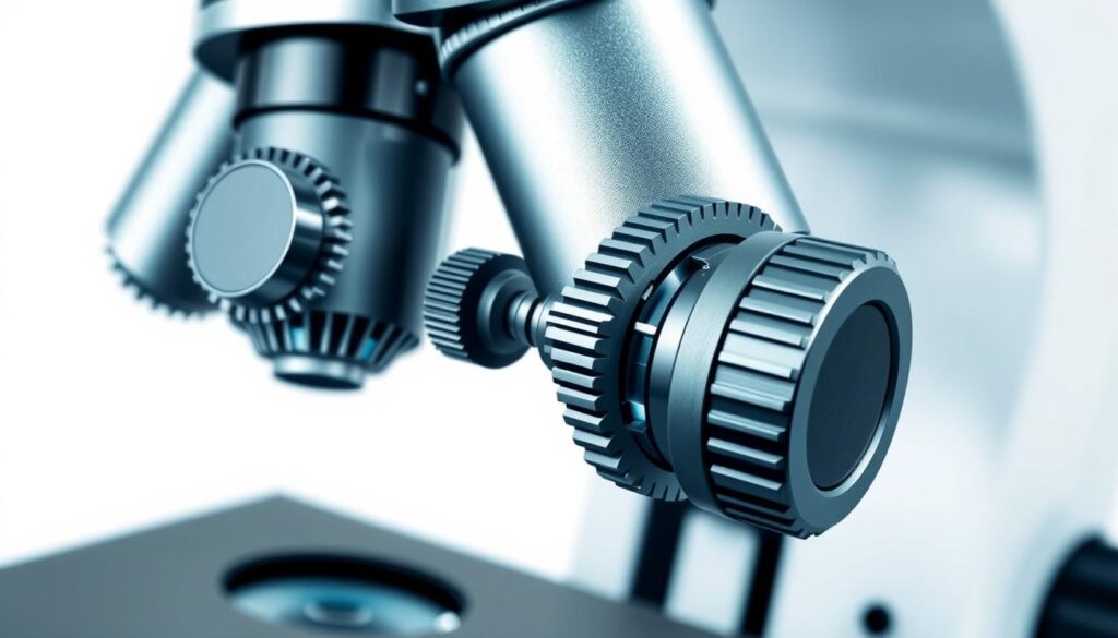
Focus Adjustment Mechanisms
Getting clear images in microscopy is key. The focus adjustment mechanisms, like the coarse and fine focus knobs, are crucial. The coarse focus knob helps with big movements, while the fine focus knob is for small, precise adjustments.
The fine focus knob moves the objective lens by just a fraction of a millimeter. This is vital for fine-tuning focus, especially with delicate specimens. Microscopes with both knobs are best for high magnifications to avoid damaging specimens.
Coarse Focus Knob
The coarse focus knob is for big adjustments and initial focusing. It works with the fine focus knob to get the focus just right.
Fine Focus Knob
The fine focus knob is for small, precise adjustments. It’s great for detailed work in research and medical diagnostics.
Working distance is also key in focus adjustment. It’s the space between the objective lens and the specimen. Adjusting focus and working distance helps get high-quality images and accurate results.
Understanding the coarse and fine focus knobs is important for microscopists. It helps them use the microscope better and reach their research goals. Proper focus adjustment is essential for clear and accurate images in microscopy.
Microscope And Its Parts With Functions: Detailed Overview
The microscope is made up of many microscope parts, each with its own functions. Knowing these parts and their roles is key to using the microscope well. The microscope’s optical system, like the eyepiece, objective lenses, and condenser lens, work together. They create a magnified image of the specimen.
Important microscope parts include the objective lenses, which can magnify from 4x to 100x. The eyepiece lenses usually have a 10x magnification power. These parts collect and focus light and adjust magnification levels. The stage and focus adjustment mechanisms are also crucial for moving specimens and getting clear images.

Modern microscopes also have advanced features like digital cameras and automated focus. These features improve the microscope’s functions and user experience. By knowing the microscope parts and their functions, users can get the most out of their microscope and achieve great results.
Light Sources and Illumination Systems
Good lighting is key in microscopy. It affects how clear the images are. Different light sources and systems are used, each with its own pros and cons. The right light source depends on the microscopy type and the goal.
Tungsten-halogen lamps, xenon arc discharge lamps, and LED lights are common in microscopy. Tungsten-halogen lamps are cheap and easy to find. But, they don’t last long and can get hot, harming the sample. Xenon arc discharge lamps last longer and give steady light but cost more.
LED vs. Halogen Options
LED lights are gaining favor in microscopy. They’re energy-saving, last a long time, and don’t get hot. They’re also better for the environment than halogen lamps. The main benefits of LED lights are:
- Energy efficiency: They use much less energy than halogen lamps.
- Long lifespan: LED lights can go up to 50,000 hours, versus 2,000 hours for halogen lamps.
- Low heat production: They don’t produce much heat, which helps keep the sample safe.
Köhler Illumination Technique
The Köhler illumination technique focuses on lighting just the area being viewed. It improves contrast by removing scattered light. This method is crucial for high-quality images in microscopy.
In conclusion, picking the right light source and system is vital in microscopy. Knowing the good and bad of each helps researchers choose the best. This leads to clearer images and more accurate results.
| Light Source | Lifespan | Heat Production |
|---|---|---|
| Tungsten-Halogen | 2,000 hours | High |
| Xenon Arc Discharge | 5,000 hours | Medium |
| LED | 50,000 hours | Low |
Understanding Magnification Power
Microscope magnification is key in microscopy. It lets us see and study things closely. The magnification power comes from the objective lens and the eyepiece. Objective lenses usually magnify from 4x to 100x. Compound microscopes can go even higher, up to 1000x or more.
To find the total magnification, you multiply the objective lens by the eyepiece. For instance, a 10x objective with a 10x eyepiece gives you 100x total magnification. Knowing how to use magnification is vital for precise results in biology, medicine, and materials science.
There are different types of microscope magnification:
- Stereo microscopes have magnification from 5x to 40x.
- Compound microscopes can go up to 1000x or more.
- Darkfield microscopy lets you see live, unstained cells and small particles.
Getting the right magnification is crucial in microscopy. It helps researchers see and study specimens well. By knowing how to use magnification, researchers can pick the right lenses for their needs. This ensures they get accurate and reliable results.
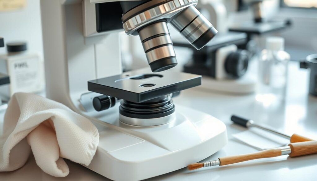
Care and Maintenance of Microscope Parts
Keeping your microscope in good shape is key to its longevity and accuracy. Regular cleaning and proper storage are vital. These steps help keep your microscope parts in top condition.
Start by wiping the microscope with a clean cloth before each use. Clean the objectives, eye-pieces, and condenser every month. For storage, use a silica packet to keep moisture away and prevent mold.
To clean the lens, mix 40% petroleum ether, 40% ethanol, and 20% ether. This blend ensures the lens works well.
Cleaning and Storage Tips
- Use a clean cloth to wipe the microscope before each use
- Clean objectives, eye-pieces, and condenser monthly
- Use a silica packet for airtight storage to absorb moisture and prevent fungal growth
- Clean the lens with a mixture of 40% petroleum ether, 40% ethanol, and 20% ether
Don’t forget about regular maintenance. Turn off the light and set the voltage to the lowest setting each day. Also, keep the microscope away from water and chemicals to avoid damage.
By sticking to these cleaning and storage tips, and keeping up with maintenance, your microscope will stay in great shape. This ensures it works accurately for a long time. Proper care is essential for your microscope’s longevity and performance.
| Cleaning Frequency | Microscope Part |
|---|---|
| Monthly | Objectives, eye-pieces, and condenser |
| After each use | Front lens of immersion objective |
Common Issues and Troubleshooting
Working with microscopes can lead to common issues that slow down research or observations. These problems can be from optical setup errors to mistakes in preparing the specimen. Troubleshooting these issues is key to avoid delays and get precise results.
Common problems include wrong lighting, incorrect condenser settings, and poor specimen prep. For example, dirt, dust, or oils on the specimen or optics can mess up one in five samples. To fix these, it’s vital to use the right troubleshootingmethods, like cleaning lenses with lens paper to avoid scratches.
- Wrong lighting and condenser settings can cause photomicrography mistakes.
- Bad specimen prep, like dirt or oils, can ruin image quality.
- Parfocal errors with low power objectives can make images blurry.
To tackle these common issues, knowing how to troubleshoot is crucial. By sticking to these methods and keeping the microscope in good shape, users can cut down on downtime and get accurate results. For example, regular cleaning of the microscope’s lenses and stages can prevent many problems and enhance image clarity.
By knowing these common issues and using the right troubleshooting steps, microscope users can improve their research and observations. This leads to more precise and dependable results.
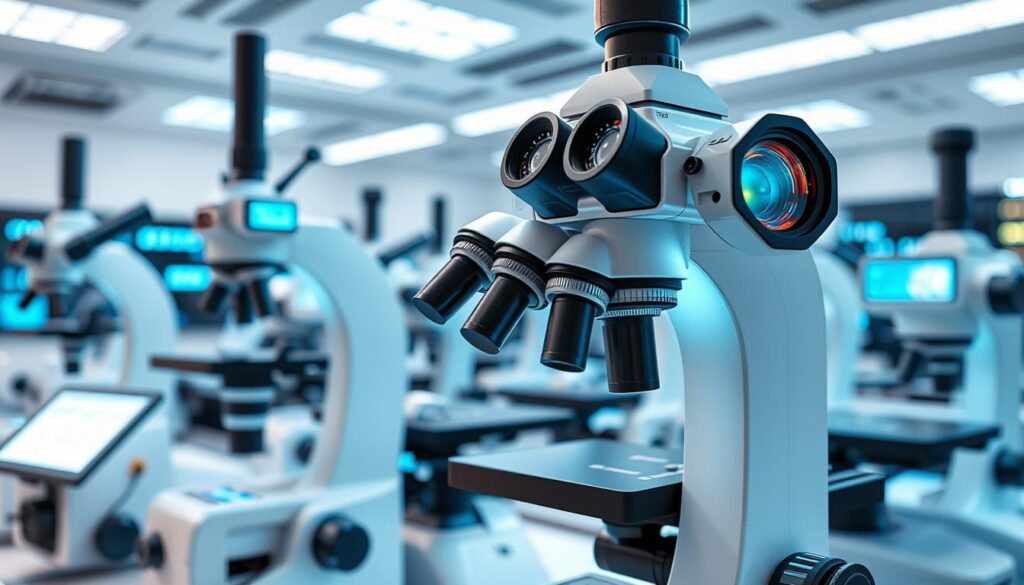
Advanced Features in Modern Microscopes
Modern microscopes have evolved a lot. They now come with advanced features that make them better. These features help in making microscopic observations more accurate and efficient. They are key in many fields of research and study.
Some important advanced features include digital integration and automated functions. Digital integration lets you capture and analyze digital images. Automated functions make adjusting settings and capturing data easier.
Digital Integration
Digital integration is a big deal in modern microscopes. It lets users take and study digital images of specimens. This feature uses advanced software to improve and change images. It helps us understand specimens better.
Automated Functions
Automated functions are another big plus in modern microscopes. They make adjusting settings and capturing data simpler. Features like automated focus adjustment and stage movement are included. This makes collecting and analyzing data easier.
Specialized Attachments
Modern microscopes also come with specialized attachments. These attachments add more functionality and versatility. Accessories like fluorescence filters and camera adapters are available. They let users customize their microscope for specific research needs.
Some examples of advanced features in modern microscopes include:
- High-resolution digital cameras for image capture and analysis
- Automated stage movement and focus adjustment for efficient data collection
- Specialized software for image enhancement and analysis
These advanced features make modern microscopes a powerful tool. They help researchers and scientists study specimens in more detail. This leads to new discoveries and a deeper understanding of the microscopic world.
| Feature | Description |
|---|---|
| Digital Integration | Enables the capture and analysis of digital images |
| Automated Functions | Streamlines the process of adjusting settings and capturing data |
| Specialized Attachments | Provides additional functionality and versatility |
Selecting the Right Microscope for Your Needs
Choosing the right microscope involves several key factors. These include your budget, the application you need it for, and your personal preferences. The right microscope can help you get accurate results and save money. First, you need to decide what type of microscope you need.
Compound microscopes are great for seeing small details, like cell structures. Stereo microscopes are better for looking at larger objects, like solder joints on circuit boards.
When picking a microscope, think about how much magnification you need. Compound microscopes can go up to 1000x, while stereo microscopes top out at 50x. Also, consider the microscope’s type – monocular, binocular, or trinocular. Trinocular microscopes are best for photos and videos because they let you use a camera without getting in the way.
Other things to think about include the type of samples you’ll be looking at and how much light you need. Digital microscopes and cameras are also important for capturing images. By considering these points, you can choose the right microscope for your needs.

Choosing the right microscope is all about understanding your needs. By carefully evaluating what you need, you can find a microscope that works well for you. Whether you’re a student, researcher, or professional, the right microscope can help you achieve your goals.
Safety Considerations and Best Practices
Working with microscopes requires careful safety measures. It’s important to handle them right and follow safety rules. This keeps everyone safe and healthy.
Keeping microscopes clean and following safety rules are key. Clean them before and after use. Also, keep them 6 feet apart to follow social distancing.
Proper Handling Techniques
Handling microscopes with care is crucial. Avoid touching the lenses and lift them correctly. This prevents damage and keeps users safe.
Laboratory Safety Guidelines
Lab safety rules are essential. Follow protocols for chemicals, wear protective gear, and know emergency steps. These steps help avoid accidents and keep the lab safe.
By focusing on safety and following best practices, we can work safely with microscopes. This includes knowing lab safety, handling techniques, and accident prevention.
- Following proper protocols for handling chemicals
- Wearing protective gear, such as gloves and goggles
- Being aware of emergency procedures, such as evacuation routes and fire extinguisher locations
- Regularly cleaning and maintaining equipment to prevent contamination
By following these tips and considering safety, we can work safely with microscopes.
| Safety Consideration | Best Practice |
|---|---|
| Proper handling techniques | Handle the microscope with care, avoid touching the lenses, and use proper lifting techniques |
| Laboratory safetyguidelines | Follow proper protocols for handling chemicals, wear protective gear, and be aware of emergency procedures |
Conclusion
Microscopes have opened up the hidden world to us. They started with Antony van Leeuwenhoek’s simple 300x microscope. Now, we have electron microscopes that can see things over a million times bigger.
The compound brightfield microscope is key in labs. It lets scientists study everything from soil to skin diseases. Techniques like fluorescent and phase-contrast microscopy give us deep insights into cells and tissues.
As we look to the future, new microscope tech will keep changing how we discover. With digital and automated features, we’ll see even more. This will let us explore the tiny world and learn more about it.
FAQ
What is the history of microscopes?
Microscopes have a rich history, starting in the 16th century. Zacharias Janssen is often credited with inventing the first compound microscope in the late 1500s.
What are the different types of microscopes available today?
Today, we have many types of microscopes. These include light, electron, and fluorescence microscopes. Each has its own special features and uses.
How do microscopes work?
Microscopes use lenses and light to show small objects in detail. They make these objects appear larger and clearer.
What are the main components of a microscope?
A microscope has several key parts. These include the eyepiece lens, objective lenses, and a condenser lens. There’s also a mirror or illuminator, stage, and focus adjustments. Each part is vital for a clear image.
How do the optical system components work together?
The eyepiece lens, objective lenses, and condenser lens work together. They create a magnified and focused image. The eyepiece lens enlarges the image, while the other lenses control light and focus.
What are the mechanical parts of a microscope and their functions?
The mechanical parts include the stage, stage clips, and focus adjustments. They hold the specimen, adjust focus, and keep the microscope stable.
How important is the base and arm of a microscope?
The base and arm are key for a microscope’s stability. The right materials and construction ensure accuracy and reliability.
How do you manipulate the stage and specimen?
To get clear images, you need to handle the stage and specimen right. Adjust the stage, use stage clips, and handle the specimen carefully.
How do the focus adjustment mechanisms work?
The focus mechanisms, like the coarse and fine focus knobs, adjust the lens to specimen distance. Proper adjustment is key for clear images.
What are the different light sources and illumination systems used in microscopy?
Microscopes use various light sources and systems. These include LED or halogen bulbs and Köhler illumination. The choice affects image quality.
How do you calculate and understand magnification power?
Magnification is crucial in microscopy. Knowing how to calculate it helps in getting accurate results and interpreting images.
How do you properly care for and maintain microscope parts?
Regular cleaning and proper storage are vital for microscope parts. This care extends the instrument’s life and ensures accurate results.
What are some common issues and troubleshooting tips for microscopes?
Microscopes can face issues like blurry images or alignment problems. Knowing how to troubleshoot can help fix these quickly.
What are the advanced features in modern microscopes?
Modern microscopes have advanced features like digital integration and automated functions. These enhance capabilities and efficiency for more complex analyses.
How do you select the right microscope for your needs?
Choosing the right microscope involves considering budget, application, and personal preferences. Making an informed decision ensures the microscope meets your needs.
What are the safety considerations and best practices for using a microscope?
Safety is crucial when using a microscope. Follow proper handling techniques and laboratory safety guidelines to ensure a safe and productive experience.